The study was performed regardless of race, sex or age and lasted 5 years during which the following protocol was used:
- Imaging examination of the upper cervical area of the patient with RX, MRI and £D CT before AtlasPROfilax® therapy.
- Evaluation of the images from various angles and exhaustive analysis of the zone C0-C1-C2.
- AtlasPROfilax® therapy
- Examination with MR and 3D CT after AtlasPROfilax® Technique.
- Comparison of the images obtained “pre” and “post” to the therapy and conclusion of each case.
The Technique was performed in the presence of the medical officers of the Radiological Diagnosticum Institute, just after the first imaging examination. A second imaging examination was then carried out under the same conditions (only about forty minutes to an hour apart from the first to the second radiological examination).
Imaging protocol
A skull carrier was attached along with a number of fastening elements. The reconstruction was graded 1/0.5mm. The cuts were performed in the sagittal and coronal planes while the axial cuts followed the orientation of the vertebral bodies, executing a simultaneous request and two acquisitions. The doses were controlled by the specialists.
The adjustment of the windows was Raw Data with HR algorithm and applying a specific convolution filter.
Results and conclusions of the study
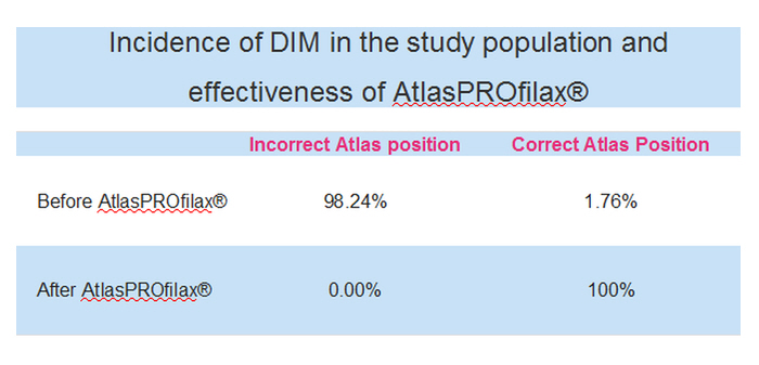
A total of 114 subjects examined 112 previously submitted to the AtlasPROfilax® technique a vicious or incorrect position of the atlas with caudal cranial deviation and inclination with significant decrease in joint contact and lack of encastramiento of the occipital condyles in the articular facets Superior of the atlas of the glenoid cavity. After the realization of AtlasPROfilax® Technique by RC Shűmperli the comparison showed an evident change in the position of the atlas. The enarthrosis of C0 with C1 was anatomically correct and the intra-articular contact between C0 and C1 as well as C1 and C2 was complete. There was also a clear improvement between the articular relation of the atlas and the axis as well as a general change of the position of the head.
It should be noted that in the vast majority of the cases examined the atlas presented an anterior left rotation in addition to an upper left tilt. The right rotation was significantly less frequent.
- Prior to AtlasPROfilax® therapy, there is more than 98 % of the cases examined with an atlas rotation with a caudal skull and tilted position (see videos and images from the study)
- After AtlasPROfilax® therapy poor atlas rotation and caudal cranial deviation with tilt are effectively corrected (see videos and study images)
- In subsequent examinations the correction of the atlas and its good position are maintained
- There results open the door to new scientific research and much broader studies not only radiological but above all on the clinical implications of AtlasPROfilax® therapy in multiple pathologies.
In addition, physicians at the Instituto Diagnosticum who performed the study did not observe adverse effects during or after the application of the AtlasPROfilax® Technique in all treated patients. The follow-up of these patients highlighted many improvements describes by themselves: improvements such as better mobility in the neck, better posture maintained over time, improvement in joint discomfort, back or limb pain, and neurovascular problems such as dizziness, poor balance or headaches.
These improvements reported by the patients have subsequently been analysed in the framework of other clinical studies.
Technology used
For the study, several technologies were used, all of them according to the latest technological standards:
- Magnetic Resonance Imaging: MAGNETOM Symphony, from Siemens.
- Computed Axial Tomography: SOMATOM Sensation 64 from Siemens, World’s fastest computed tomography scanner (2004-2006). The SOMATOM Sensation 64 sets new standards not only for speed but also for image quality and details.
Incidence of DIM in the study population and effectiveness of AtlasPROfilax®


